
The pig's respiratory system and its defense mechanisms






What kind of cells make up the respiratory defense system of pigs? How do they organize and communicate with each other?
 3tres3.com
Professional Pig Community
3tres3.com
Professional Pig Community


The pig sector events all around the world

Dernières nouvelles de la filière porcine
Un résumé tous les 15 jours des nouveautés de 3tres3.com
Information on pig health: news and articles, disease guide, diagnostic tool, clinical cases, atlas of pathology…

Simulator that calculates the amount of drug to add to the water when using a flow dispenser.

Definition for the most commonly used pig terms

Description of the most important diseases and conditions in pigs

Pig disease diagnostic tool

Images of major swine diseases

All the information about ASF: how to recognize the disease, how it is transmitted, pictures of lesions, latest news, guides, etc.

A visual and practical step-by-step guide on how to perform a necropsy on a pig.

Biocheck.UGent is an independent, risk-based, scientific scoring system for assessing the quality of your on-farm biosecurity.

All the information on Foot and Mouth Disease in pigs: how to recognize the disease, how it is transmitted, images of lesions, latest news, guides,...
Un résumé tous les 15 jours des nouveautés de 3tres3.com
Articles on nutrition and pig feeding, characteristics of raw materials and additives for pig feed. Prices of raw materials
Definition for the most commonly used pig terms

Technical sheets of the main raw materials and additives used in swine feed. They include a comparison of nutritional values from various sources, product

Global production and trade data for the most important raw materials

Latest quotations for the main commodities used in pig feed. Historical graphs with the pig price and estimated feed price.
Un résumé tous les 15 jours des nouveautés de 3tres3.com
Articles on genetic improvement and pig reproduction
Definition for the most commonly used pig terms

Compare production data, calculate the number of sow, nursery, and finishing spaces, and visualize your tasks on the work schedule by type of BMS.

Tool that allows you to calculate the replacement rate in your farm
Un résumé tous les 15 jours des nouveautés de 3tres3.com
Information on the design of pig farm facilities, equipment and farm
Simulator that calculates the amount of drug to add to the water when using a flow dispenser.
Definition for the most commonly used pig terms
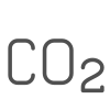
Environmental Footprint Calculator along the pork value chain.
Biocheck.UGent is an independent, risk-based, scientific scoring system for assessing the quality of your on-farm biosecurity.
Un résumé tous les 15 jours des nouveautés de 3tres3.com
News and economic comments on the market situation of pork and raw materials. Prices of pig and raw materials. Census, pork production and trade data

The latest slaughter pig prices in the most important pig markets. Check the evolution of the historical prices in charts and in several currencies.

Figures & trends in pig numbers, pork production and pork trade.
Global production and trade data for the most important raw materials
Latest quotations for the main commodities used in pig feed. Historical graphs with the pig price and estimated feed price.
Un résumé tous les 15 jours des nouveautés de 3tres3.com
Management, pig farm management, work planning in each production stage
Definition for the most commonly used pig terms
Compare production data, calculate the number of sow, nursery, and finishing spaces, and visualize your tasks on the work schedule by type of BMS.
Tool that allows you to calculate the replacement rate in your farm
Un résumé tous les 15 jours des nouveautés de 3tres3.com


What kind of cells make up the respiratory defense system of pigs? How do they organize and communicate with each other?

A discussion of the different micro and macroscopic lesions caused by PRRSV, PCV-2, swine flu virus, and porcine respiratory coronavirus.

This article describes the principal lesions that characterize the main bacterial pneumonias. Although most are opportunistic agents, there are two primary bacterial agents capable of causing lesions on their own.

M. hyopneumoniae is involved in most of the co-infections of the porcine respiratory system. How does it act and what effects does it have?

What is the actual effect of pneumonia on the farm's economic performance? What similarity is there between pneumonia and heat stress?

This new section, coordinated by the pathological anatomy team of the University of Cordoba, Spain, will show us, in a very visual and practical way, how to perform a necropsy on a pig.

In this second installment, we cover extracting the tongue, trachea, lungs, and heart in one piece. We will extract the organs of the abdominal cavity, both the digestive system and the urinary and genital tracts. In the head, we will access the nasal turbinates, brain, and cerebellum.

In the first two installments, we covered necropsy in lateral decubitus. In this third installment, we will look at necropsy in the supine position

In the fourth installment of the manual, we address examining the thoracic cavity organs.

In the fifth installment of the manual we address the examination of the organs of the abdominal cavity.

After interpreting the lesions we must proceed to sample collection.

To complement the necropsy course, we have produced a series of videos of different processes in pigs.